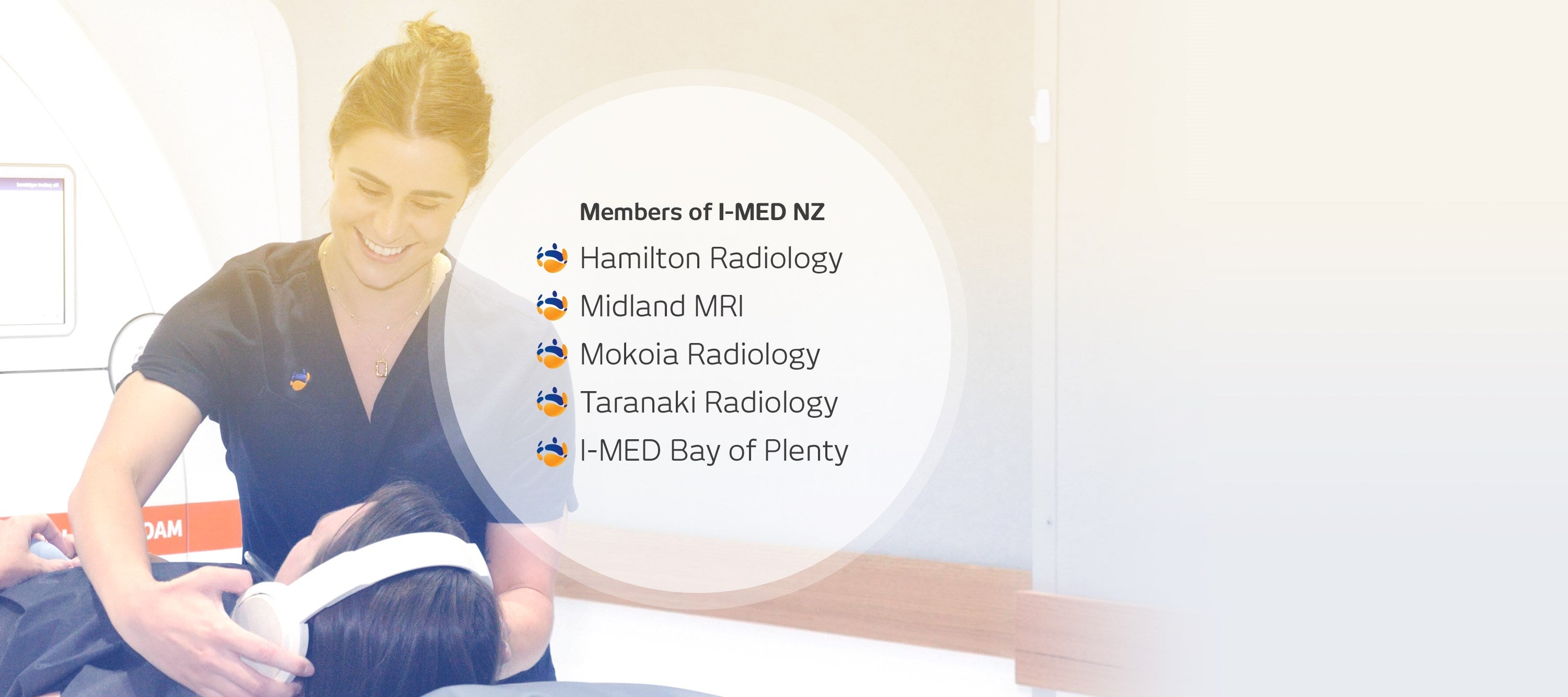
I-MED Radiology NZ, when you need answers
With 23 clinics across the North Island and a team of 30 experienced radiologists renowned for their subspecialist expertise, I-MED Radiology NZ is committed to exceptional patient care. Our advanced technology can provide accurate diagnoses, for a clear picture of your health.
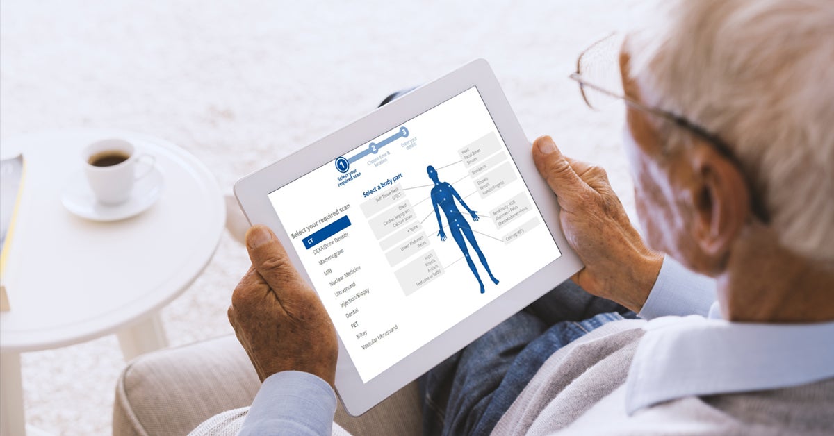


Our process is simple and patient focused
2
Prepare and attend
Depending on the procedure, you may be asked to fast or drink water beforehand. It’s important to read through the procedure information, prepare appropriately before your appointment, and arrive for your appointment in plenty of time.
3
Speak with your doctor
Once our radiologist has finalised your results, I-MED will send a report directly to your referring practitioner. It is important to speak with your health practitioner about your result, to understand what they mean and complete follow up treatment if needed.
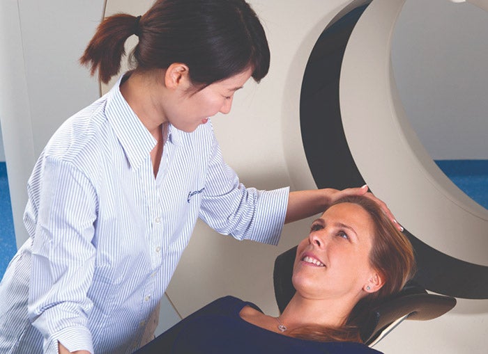
Our clinics offer a range of medical procedures
I-MED Radiology clinics offer a range of imaging procedures including MRI, CT, X-ray, Ultrasound, PET/CT and Interventional Services. We take pride in our commitment to employing the most up-to-date equipment, as well as highly qualified and specialised Radiologists and staff across all modalities.