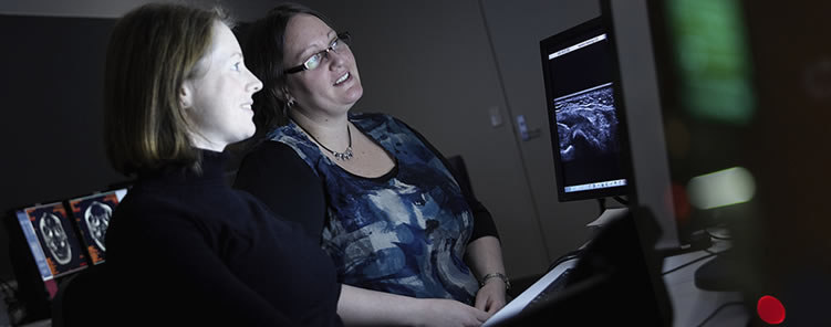
Hamilton Radiology provide a wide range of Ultrasound services across the Waikato region. Follow the links below to learn more about each of these.
What is ultrasound?
Ultrasound uses high frequency sound waves to demonstrate the internal structures of the body. The echoes of the sound waves are recorded and displayed as a visual image. Ultrasound cannot be used for all areas of the body - some structures/areas may be better seen with X-rays, CT or MRI.
What is ultrasound used for?
An ultrasound of the abdomen (stomach area) is a useful way of examining the internal organs (e.g. liver, gallbladder, kidneys, and bladder). Ultrasound images are shown on the screen and can demonstrate blood flow in some studies (see below).
Doppler ultrasound
Doppler ultrasound enables us to look at the circulatory system (blood in arteries and veins) Doppler ultrasound is used to diagnose any problems such as blockages or narrowing of the blood vessels. The blood flow is demonstrated on the ultrasound picture as areas of differing colours.
What do I need to do before an ultrasound?
You may need to remove clothing (e.g. for an ultrasound of your abdomen, we need to be able to see all of your abdomen, so it can be helpful if you are wearing clothes that are easy to slip up or off).
Some of the ultrasound examinations require that you either drink water, or alternatively don't eat or drink. The instructions will be given when making your appointment and/or an instruction leaflet will be sent to you along with a confirmation of appointment letter. Where relevant, instructions are also on this website.
What does the ultrasound room look like?
The ultrasound room is darkened and has a narrow bed, beside which there is a machine with a monitor much like a TV or computer screen. The front of the machine looks like a computer keyboard. The part of the machine that is used to produce the ultrasound image is called the "transducer" This is a small plastic square attached to the ultrasound machine by a thick cord.
When you have the ultrasound examination, the Sonographer (technologist who is trained in ultrasound) will spread gel on the area to be examined. This ultrasound gel enables good contact between the transducer and the skin so the best images are obtained. The Sonographer may ask you to move around and/or to hold your breath so they can see the area to be examined clearly.
The Sonographer will watch the monitor during your examination, and may point out fetal anatomy on pregnancy scans.
The Sonographer may ask you to wait in the ultrasound room until they have shown the ultrasound images to the Radiologist. The Radiologist may wish to take some further ultrasound images. This does not mean something is wrong, rather that they may wish to see the moving images of an area.
After the examination
The Radiologist will review the images and provide a written report to your referring doctor. Please settle your account on the day of the examination.
Please contact Hamilton Radiology for an appointment on 07-839 4909 or 0800 HAMRAD (0800 426723)